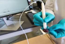
Ten seconds is all it could take to detect cancerous tissue during surgery, thanks to a novel device that researchers believe has the potential to transform cancer treatment.

In a new study, scientists reveal how the device, which is called the MasSpec Pen, was highly accurate in detecting cancer in human tissue samples, and it did so in just 10 seconds.
Study leader Livia Schiavinato Eberlin, of the University of Texas at Austin, and colleagues say that the tool could vastly improve the accuracy of cancer surgery and help to reduce recurrence of the disease.
The researchers recently reported their findings in the journal Science Translational Medicine.
Last year, more than 1.6 million new cancer cases were diagnosed in the United States, and more than 595,000 people died from the disease, making it one of the leading causes of death in the country.
Surgery remains one of the primary diagnostic and treatment strategies for cancer. It aims to detect and remove cancerous tissue and prevent it from spreading to other parts of the body.
However, distinguishing between healthy and cancerous tissue can prove tricky for surgeons, making it difficult for them to remove all cancer remnants.
Frozen section analysis - also referred to as cryosection - is one technique that aims to help with this problem. This involves taking a tissue sample from a cancer patient during surgery and transferring it to a laboratory for freezing, where it is then assessed by a pathologist.
But Eberlin and colleagues say that this method can be slow, which may increase a patient's risk of surgery-related complications. Furthermore, they note that frozen section analysis can be unreliable for some cancer types.
How does the MasSpec Pen work?

The team believes that the MasSpec Pen could offer faster, more accurate detection of cancerous tissue during surgery.
The state-of-the-art device works by identifying tissue metabolites that are unique to cancer cells, using a technique called mass spectrometry.
"Cancer cells have dysregulated metabolism as they're growing out of control. Because the metabolites in cancer and normal cells are so different, we extract and analyze them with the MasSpec Pen to obtain a molecular fingerprint of the tissue," explains Eberlin.
The molecular fingerprint that has been drawn from tissue is then assessed using "statistical classifier" software.
The team "trained" this software to distinguish between cancerous and non-cancerous molecular fingerprints by feeding it data from hundreds of healthy and cancerous human tissue samples, including tissue from the lung, breast, and ovary.
Once the device has assessed the tissue, it will flag the words "Normal" or "Cancer" on a computer screen.
Wanna know more?? Write us @ surgicalnursing@nursingmeetings.com
*Reference article: MEDICAL NEWS TODAY
No comments:
Post a Comment